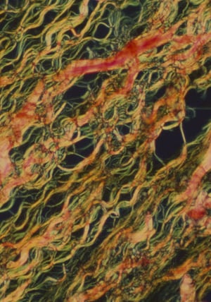

Collagen fibers stained with Picrosirius Red allows distinction between two fiber types using polarized light observation
To observe collagen fibers, samples can be stained with Masson trichrome (MT), Elastica van Gieson (EVG), or Elastica Masson (E-M); however, distinction between collagen fiber types I and III is not possible with this method. In drug discovery research, quantitative evaluation of the collagen fibers is required, so it is necessary to be able to distinguish between collagen fiber types I and III.
Download the app note to learn more about:
Download the application note, courtesy of Olympus